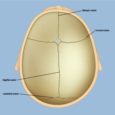
A misshapen head in a baby may be the result of craniosynostosis, which is a deformity of the skull caused by premature fusion of growth plates of the skull, called sutures. Craniosynostosis occurs in one out of every 2,000 live births and is more common in boys than girls.
Visit our Craniosynostosis program page
Infants and children with craniosynostosis experience progressive abnormal head shape and ultimately, if the condition is left untreated, an increase in intracranial pressure, which can affect development. The condition can be safely corrected by an experienced pediatric craniofacial neurosurgeon. Depending on the form of synostosis a child has, the surgical team may also include a plastic or reconstructive surgeon (see Doctors Who Treat Craniosynostosis).
Note that craniosynostosis is different from the “flat head” that sometimes occurs in babies who spend a lot of time on their backs — a condition called deformational plagiocephaly, or positional molding. Deformational plagiocephaly is self-correcting and resolves on its own over time, but craniosynostosis does not.
A Guide for Physicians: Diagnosing and Treating Craniosynostosis
A Parent's Guide to Craniosynostosis Surgery
The skull is not one large bone — it consists of several bones that are held together by fibrous growth plates that allow the skull to expand as the infant’s brain grows and develops. These fibrous openings, known as sutures, normally begin to fuse (or close up) starting at a few months of age and continuing to approximately 3 years, depending on the suture, after the rapid brain growth that occurs during an infant’s first 36 months slows down. In some children, however, one or more of the sutures can fuse early; because the brain is still growing at its normal rate, the premature closure of the suture results in an abnormally shaped head.
There are several types of craniosynostosis, depending on which suture is involved, and each type creates a distinct head shape (see Symptoms of Craniosynostosis). Some rare cases of craniosynostosis may be part of a larger syndrome, but the overwhelming majority are isolated (also called nonsyndromic), meaning that only one suture is involved and no other part of the body is affected. These isolated conditions include:
Sagittal synostosis is the most common form of craniosynostosis; it is caused by the premature fusion of the sagittal suture, which runs front to back along the middle of the skull, separating the left and right portions of the skull. The long, narrow skull shape that results is known as scaphocephaly, often referred to as a “boat shape.”
Unilateral or bilateral coronal synostosis is the second most common form of craniosynostosis. It is caused by fusion of the coronal suture, which runs crosswise on the top of the skull (from ear to ear) and is divided in half by the sagittal suture. In unilateral coronal synostosis, either the left or right sided coronal suture closes prematurely; you may notice that one eye is slightly higher than the other, that one ear is further forward than the other, or that the nose appears tilted. In bilateral coronal synostosis (which is less common), both sides of the coronal suture fuse too early.
Metopic synostosis is caused by the fusion of the metopic suture, which runs from the top of the skull down the center of the forehead to the nose. A child with metopic synostosis may have a triangular-shaped head, known as trigonocephaly.
Lambdoid synostosis is caused by the fusion of the lambdoid suture, located on the back of the skull and shaped like an upside down “V.” Usually only one side fuses, but there have been rare cases in which both sides fuse. A child with lambdoid synostosis may appear to have one side of the head flatter than the other, a low bump behind the ear on the affected side, and a “windswept” appearance of the other side of the skull.
See illustrations of all types of craniosynostosis on the Symptoms of Craniosynostosis page.
What Causes Craniosynostosis?
It is not clear why some children experience a premature fusion of the sutures. In some cases there is a family history of the abnormality, but more often there is no apparent cause. A child with craniosynostosis usually has no other abnormality, but in some cases it is part of a larger syndrome caused by a genetic mutation. Children with craniosynostosis should be examined to rule out other possible genetic disorders or malformations.
Children with craniosynostosis or other craniofacial abnormalities are best treated at a major medical center with a comprehensive Craniofacial Program, where experts from a wide range of disciplines have expertise in craniofacial disorders.
Request an Appointment | Refer a Patient
Reviewed by: Caitlin Hoffman, M.D.
Last reviewed/Last updated: June 2023
Illustrations by Thom Graves, CMI