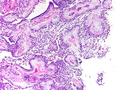
Pictures of Myxopapillary Ependymoma

These images, courtesy Dr. David Pisapia, show ependymoma cells that have been stained with hematoxylin and eosin (H&E). The stained sections demonstrate hyalinized vessels within a basophilic, myxoid material that is surrounded by arrays of neoplastic glial cells in a papillary architectural configuration. 
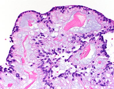
Ependymoma - courtesy  David Pisapia, MD


Ependymoma - courtesy David Pisapia, MD

Our Care Team

  • Chair and Neurosurgeon-in-Chief
  • Margaret and Robert J. Hariri, MD ’87, PhD ’87 Professor of Neurological Surgery
Phone: 212-746-4684
  • Chief of Neurological Surgery, NewYork-Presbyterian Brooklyn Methodist
  • Professor, Neurological Surgery
  • Director, Brain Metastases Program
  • Co-director, William Rhodes and Louise Tilzer-Rhodes Center for Glioblastoma
Phone: 212-746-1996 (Manhattan) / 718-780-3070 (Brooklyn)
  • Associate Professor, Neurological Surgery
Phone: 718-670-1837
  • Professor of Clinical Neurological Surgery
Phone: 212-746-2438
  • Chief of Neurological Surgery, NewYork-Presbyterian Queens
  • Professor of Clinical Neurological Surgery
  • Co-director, Weill Cornell Medicine CSF Leak Program
Phone: (718) 670-1837

Weill Cornell Medicine Neurological Surgery 525 East 68 Street, Box 99 New York, NY 10065 Phone: 866-426-7787