Surgery to remove an orbital tumor is complex due to its delicate location and should only be performed by highly trained neurosurgical teams with experience in the procedure. The goals of the surgery are:
In some cases, the only way to remove the tumor and protect the patient’s life is to perform an enucleation, or removal of the eye. Fortunately, this can often be avoided when surgery to remove the tumor is performed by a highly skilled team that includes a neurosurgeon, a plastic surgeon, and an ophthalmic surgeon. When enucleation is required, a prosthetic eye can be attached to the muscles so that it moves in synch with the remaining eye, for good cosmetic results.
The Surgical Approach to an Orbital Tumor
One of the difficulties in surgery for an orbital tumor is determining the approach, or the path the surgeon or surgeons will take to reach the tumor. (In major medical centers, the surgery is often performed by both a neurosurgeon and ophthalmologic or oculoplastic surgeon working together.) Depending on the location and nature of the tumor, the surgeons may be able to approach from the side (laterally), or from above or below the eye (with incisions along the eyebrow or eyelid).
The Transorbital Approach
In many cases, the approach can be done through a small incision in the eyelid using an endoscope. Weill Cornell Medicine neurosurgeons, working together with oculoplastic surgeons, have pioneered this new minimally invasive approach and are world leaders in this type of surgery. In general, the transorbital approach leads to faster recovery and a better cosmetic outcome since the incision is much small than a more lateral approach through the side of the head.
Tumor Removal and Skull Reconstruction
Surgery to remove an orbital tumor may last between four and eight hours, depending on the size and complexity of the lesion. Reconstruction of the skull and/or orbit (eye socket) may be required as part of the treatment. Additionally, acquired defects caused by trauma or benign lesions such as Graves’ disease and Paget’s disease may also require reconstructive treatment. Although the patient’s own body tissue is usually best in any reconstruction, advances in bone and surgical technology have yielded new and exciting materials that may serve as a substitute when needed. These advances include titanium plates and screws, hydroxyapatite cement, porous polyethylene, and resorbable fixation devices.
Recovery
Patients are usually hospitalized for two to seven days, and recovery time can be from two to six weeks.
Surgery for Graves’ Disease
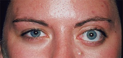
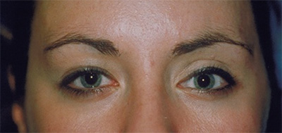
Some patients with Graves’ disease require a surgical procedure to reposition the eyelids. In more severe cases the eye itself and/or the optic nerve may require repositioning or decompression. A skilled surgeon can make changes in the bone around the eye (orbit) that achieve excellent cosmetic and functional results.
Unilateral Graves: Orbital Bone Repositioning
Pre-Op
Late Post-Op
Reprinted with permission from: Spinelli, H.M., Atlas of Aesthetic Eyelid and Periocular Surgery. Philadelphia: Saunders-An Imprint of Elsevier, Inc., 2004.
Reviewed by: Theodore Schwartz, MD
Last reviewed/last updated: June 2024