It's only natural to want to know what a Chiari malformation looks like. These images will help you understand what a Chiari malformation is, and how decompression surgery helps to resolve it.
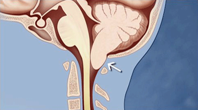
This illustration shows the cerebellar tonsils descending from the skull toward the spinal column, creating pressure.
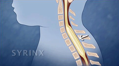
This illustration shows a syrinx, which is a cyst in the spinal column.
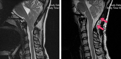
These MRI scans show a patient before (left) and after (right) surgery for Chiari malformation. The scan on the right shows that the cerebellum has returned to a normal position, and the red arrows show how the cerebrospinal fluid surrounding the cerebellum has returned to normal.
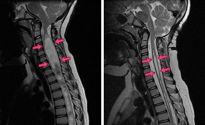
These MRI scans show a Chiari patient's syrinx (left, before surgery), and the reduction in the size of the syrinx after surgery (right, arrows). After the surgery, cerebrospinal fluid surrounding the cerebellum also returns to normal.
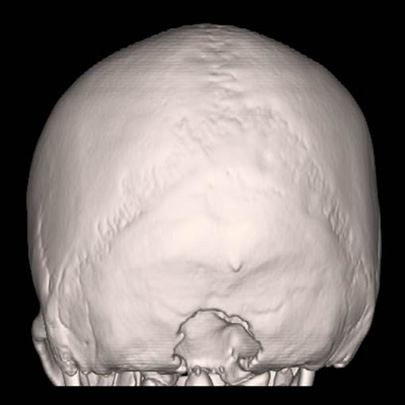
Taken after minimally invasive decompression surgery, this CT scan shows the area where bone was removed.