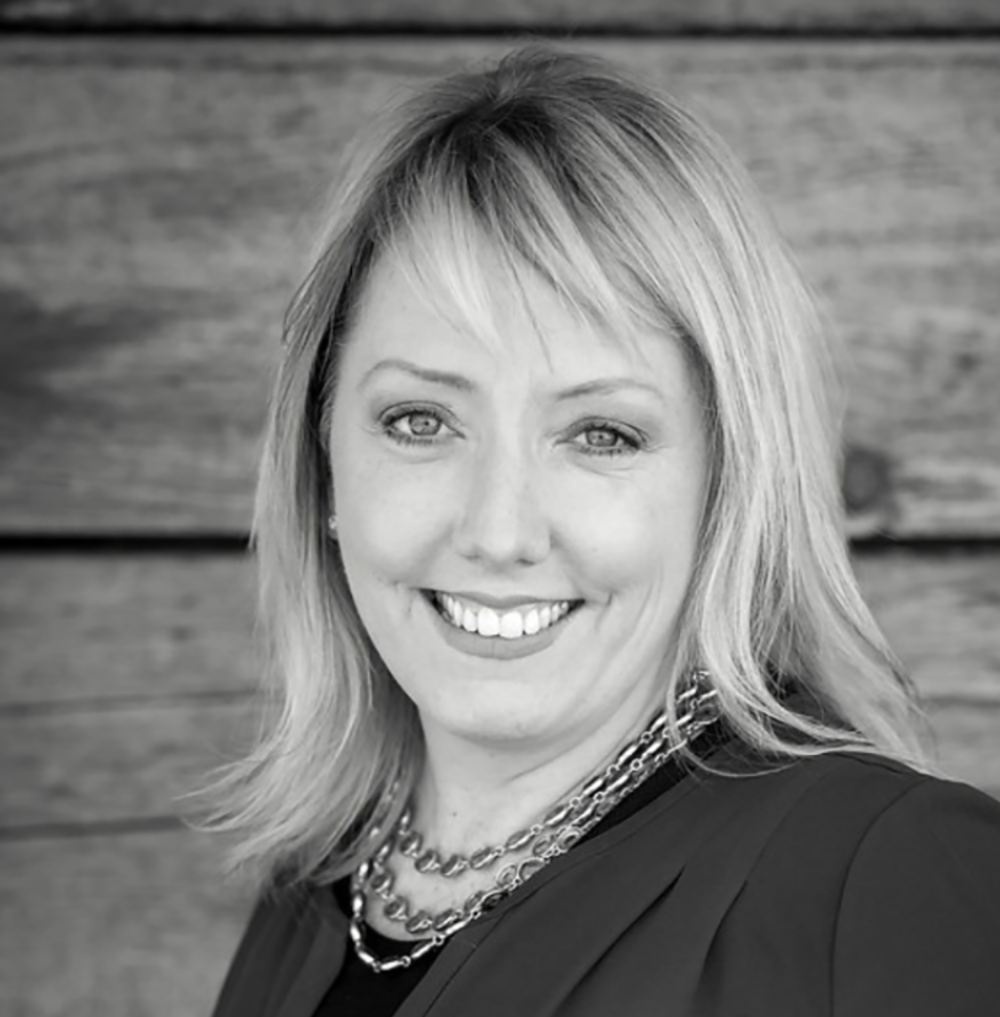
It started out like any other normal day for Keri Mahe, a 40-year-old mother of two from Erie, Colorado. After her daily Spin class, she dropped her kids off at school and preschool, then stopped by a local coffee shop before settling down to work. But after picking up her latte, she noticed something strange — she couldn’t feel the warmth of the cup in her hand. Then the left side of her face and body started tingling. “I knew instinctively that something was wrong,” she recalls. “And I knew it was something neurological.”
Keri quickly texted her husband, Fred, to tell him that she was heading to the nearest hospital. There, a MRI of her brain showed that Keri had had a hemorrhagic stroke. Bleeding had occurred in her brainstem due to a cavernous malformation — an abnormal cluster of small blood vessels. Keri was shocked. “I had never had any medical issues in my life,” she said. “And I had never heard of a cavernous malformation.”
Cavernous malformation (CM, often referred to as a cav mal) is a tangle of tiny blood vessels and larger, thin-walled vein that can periodically bleed. A CM can be hereditary and congenital, starting as a brain lesion before developing into a malformation. Luckily, Keri’s stroke seemed to have caused no lasting damage. “The doctors eventually sent me home,” she said, “but they urged me to see a neurosurgeon.”
Later that week, a local neurosurgeon confirmed that Keri’s cavernous malformation had bled – and might bleed again. But removing it was not something he recommended. “It was in an area of my brain where the risks of removing it far outweighed the benefits,” she says. Her CM was deep within the pons of her brainstem, a major structure that controls the flow of information between the brain and the rest of the body and controls many important bodily functions, like balance and hearing.
Keri wanted a second opinion from a neurosurgeon who specialized in CMs. Having grown up in the New York City area, she called family and friends there for recommendations. “I was referred to Dr. Jared Knopman at Weill Cornell Medicine and was told he was the best.”
Keri flew to New York for a consultation. “Dr. Knopman immediately made me feel like I was in incredible care,” she says. “His extensive knowledge and experience with cavernous malformations was impressive.” Dr. Knopman agreed that because of the CM’s location, it was better to do nothing. “He said it was in a ‘high rent’ area — literally sitting on everything the brain controls — and the risk of surgery was too great,” recalls Keri. “He also said cavernous malformations behave differently in everyone and there was a chance that the CM would never bleed again.” He suggested monitoring it to determine if it had a favorable natural history, and Keri returned to Colorado comforted, but concerned. “It was good to know that both Dr. Knopman and my local doctor agreed on next steps,” she says. “Still, I felt like I had a ticking time bomb in my head and worried that it would hemorrhage again.”
Unfortunately, it did. Six weeks after her first stroke, Keri had another one. This time, the bleeding left her with balance issues and crippling neuro-fatigue. A new MRI was sent to Dr. Knopman, who called Keri with an updated recommendation. Based on the evidence that her cavernous malformation was an active type, he encouraged her to have surgery to remove the malformation. “He explained that any time the CM bled and rebled, the neurological deficits I would experience would be greater, and I’d have a harder time bouncing back over time,” she said. “And I could, God forbid, face death as well.”
But there was a glimmer of good news. In the new scan, Dr. Knopman noticed that the malformation had moved closer to the edge of the brainstem, where it could more safely be removed. Still, he warned, the surgery would be risky and her recovery would be tough. Because the malformation was in an area rich in the neural connections that control balance, coordination, speech, vision, and facial movement, surgery would likely result in neurological deficits — some that might resolve themselves on their own over time, some that might not. Keri would need help regaining function post-surgery through physical and occupational therapy.

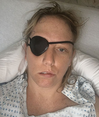
Keri Mahe wore an eye patch at first after surgery to repair her cavernous malformation
“He was very direct and up front about what to expect after coming out of surgery,” says Keri. “I’d have a hard time walking, my face would be paralyzed, I’d have double vision — it could take eight months to a year to recover. He told me that I’d need mental strength to get through it.”
But Dr. Knopman was encouraging. Keri was young and fit and had a long life ahead of her. “He said, ‘It’s going to be hard, but look at it this way — you can take your licks up front or take them over the long haul,’” Keri recalls. “I decided that I wanted to get it over with and take them all up front.” Bracing herself for what she knew would be the hardest year of her life, she agreed to have surgery.
A craniotomy was scheduled for March 15, 2017. Keri’s husband and parents fought back tears of worry as she was wheeled into surgery, but Keri felt surprisingly calm. “Dr. Knopman and his team told me exactly what they were doing and why, so I never felt confused or scared. I was informed, prepared, and cared for in the best way. And I was completely confident in a positive outcome,” she says. “The communication was critical to my overall feeling of assurance and peace that day.”
The procedure was a success. During the four-and-a-half-hour operation, Dr. Knopman and his team carefully opened her skull and removed all the diseased blood vessels, while being as gentle to surrounding brain as possible.
Waking up after surgery, Keri felt everything Dr. Knopman told her to expect — pain, numbness, double vision, facial paralysis, fatigue, jaw tightness, and tinnitus. “He told me that I’d feel like I’d been hit by a bus, and I did,” she recalls. “I wasn’t shocked by any of it.”
After spending five days in the hospital, she went home to her parents’ house in New Jersey to start her long recovery, while Fred and her older child, Dylan, returned to Colorado (her younger child, Caroline, stayed with her). For the next month, Keri underwent physical and occupational therapy to help her walk, talk, and eat normally again. Humbled by having to use a walker, she gradually willed herself to walk on her own. Her double vision went away and the pain subsided, though she still had some facial paralysis and had to wear a patch over one eye because it wouldn’t fully close.

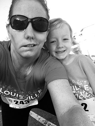
Keri Mahe and her son, Dylan, participated in a 5K run just two and a half months after her brain surgery
One month after surgery, Keri returned to Colorado and continued her recovery, happy to be reunited with her husband and son. She started running again — slowly working her way back up to three to five miles each week. Still, she was extremely tired and frustrated by ongoing balance and numbness issues. Despite ongoing support from family and friends, Keri felt alone and overwhelmed.
“I needed to talk to people who had been through what I was going through,” she says. “So I reached out to others who had experienced strokes through various organizations and support groups. This was invaluable and a critical point in my healing process.”
Keri took comfort in talking to other survivors and sharing her story. And slowly, over time, life got easier. Today, nine months after surgery, her energy is back. She’s working full-time again and has returned to her daily Spin classes. Her facial nerves have healed, though her face still droops on one side, which she’s planning to correct through reconstructive surgery this spring. “I am ready to smile again and laugh,” she says.
Dr. Knopman couldn’t be more pleased with her recovery. At her recent checkup, he said her MRI looked great. Best of all, the risk of having another bleed and stroke are gone. “I feel like I’ve been given a second and third chance at life, with a long runway to make things even better than they were before,” says Keri. “I have seen the best in my family and friends though this experience — and the best in myself.”
More about cavernous malformations
More about Dr. Knopman
More about our Stroke Program