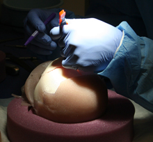
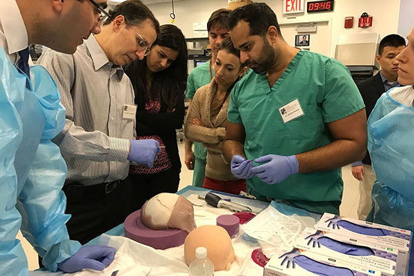
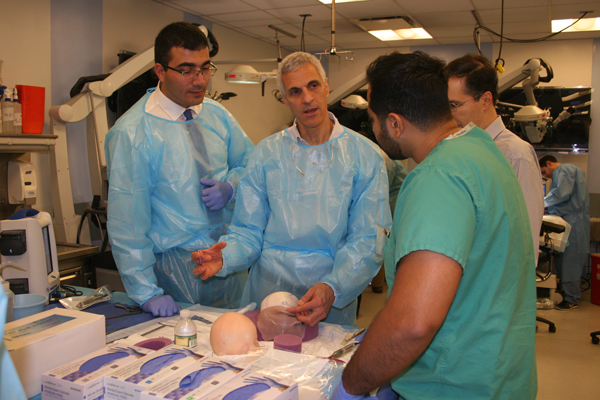
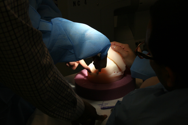
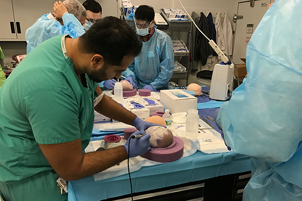
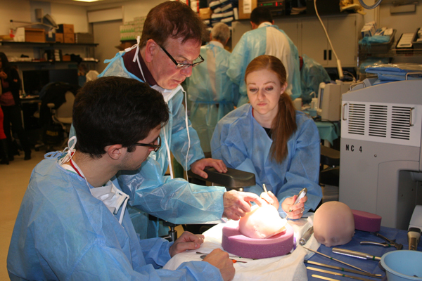
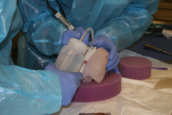
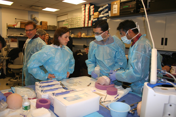
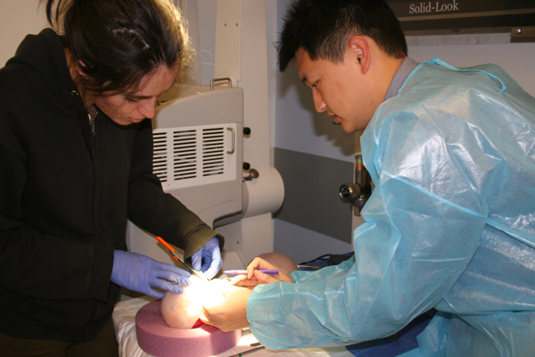
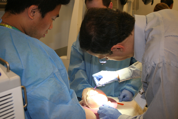
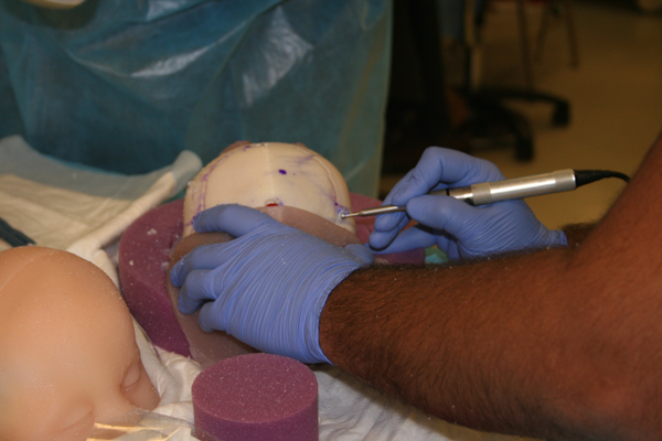
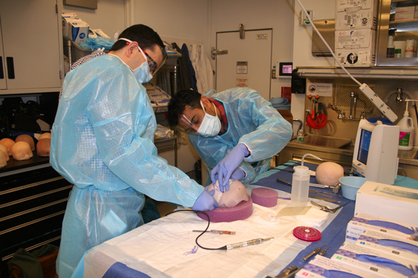
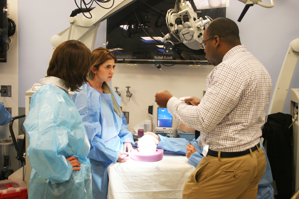
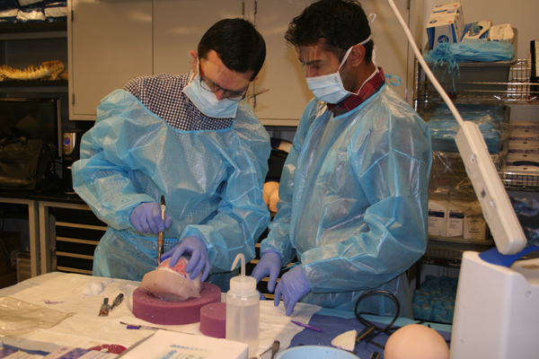
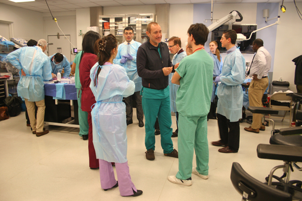
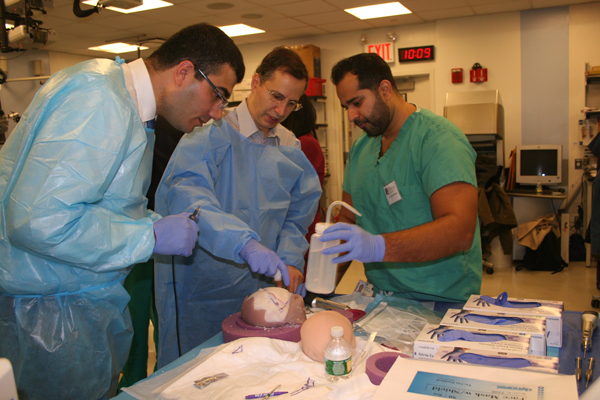
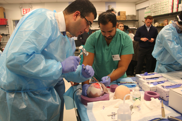
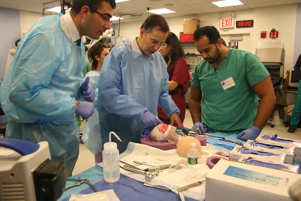
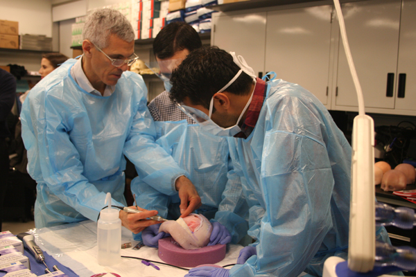
A recent training course at Weill Cornell Medical College, co-directed by neurosurgeon Mark Souweidane of Weill Cornell Medicine and plastic surgeon Jeffrey Ascherman of Columbia University, provided an innovative opportunity for young surgeons to acquire hands-on experience repairing craniosynostosis using unique 3-D printed models.
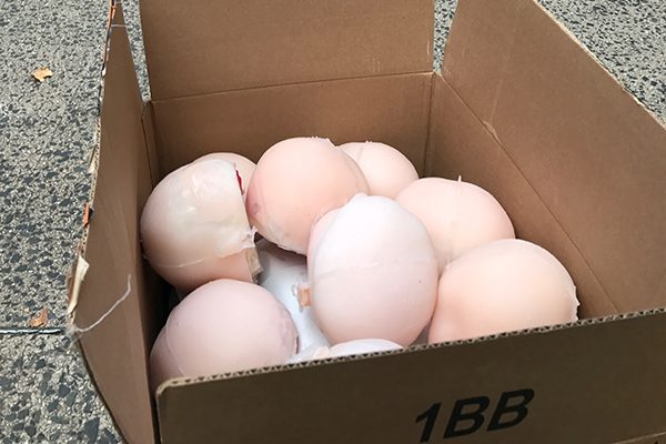
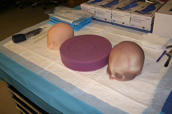
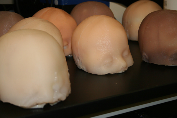
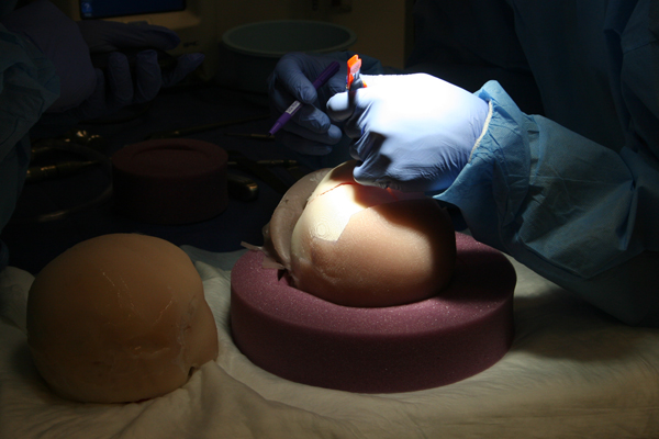
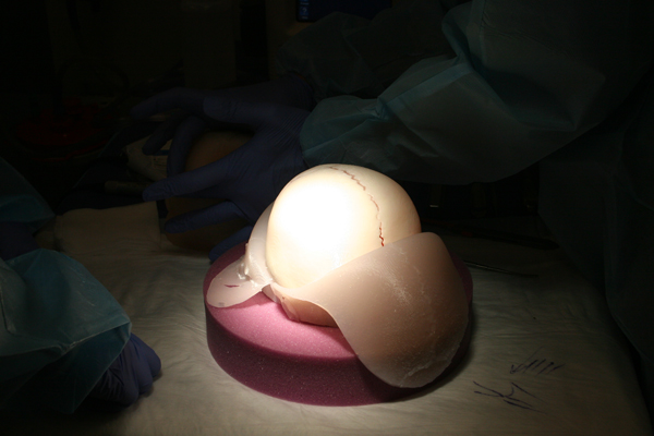
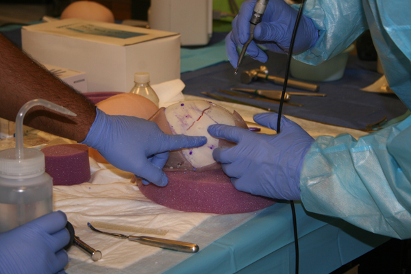
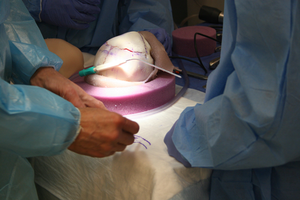
The models were created by Du Cheng, a tri-institutional (Weill Cornell Medical College, Memorial Sloan Kettering, and Rockefeller University) MD/PhD candidate, who has been working on new ways to use 3-D printing to create models of infant heads based on actual patient scans. These intricate models include realistic layers of skin, bone, dura, and brain, allowing trainees to learn and practice the remodeling surgery that these infants require.
These models fill a great need, since remodeling surgery to repair craniosynostosis is a complicated one and it has been difficult to train neurosurgeons and plastic surgeons to develop expertise in it. Not only is the condition relatively uncommon, there is also no ability for surgeons to train on cadavers as they usually do. This innovation in 3-D printing promises to bring new opportunities for experts to teach these advanced techniques to more young surgeons. The December 2016 course, held in the state-of-the-art Surgical Innovations Laboratory at Weill Cornell Medical College, allowed more than a dozen young neurosurgeons and plastic surgeons to train under a faculty of experts using these new models. (Article continues after slide show.)
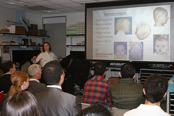
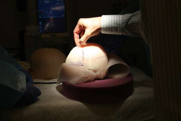
Approximately one in every 2,000 infants is diagnosed with craniosynostosis, which refers to the premature fusion of bony plates that are meant to keep expanding until the child’s rapidly growing brain reaches its full size. The brain continues to grow, but since it can’t expand properly in all directions the child’s head takes on a distorted appearance – if the prematurely fused suture runs from front to back, for example, the skull cannot expand at the sides and thus expands abnormally front to back, causing an elongated appearance. (See more about craniosynostosis.)
Cranial vault remodeling surgery reopens the prematurely fused sutures, allowing the skull to continue its normal expansion and repairing the distortion in the head shape. The ability of experienced surgeons to use these realistic models will provide an unprecedented opportunity for them to train their young colleagues in this complex procedure.